Contact Details
Clinic Hours
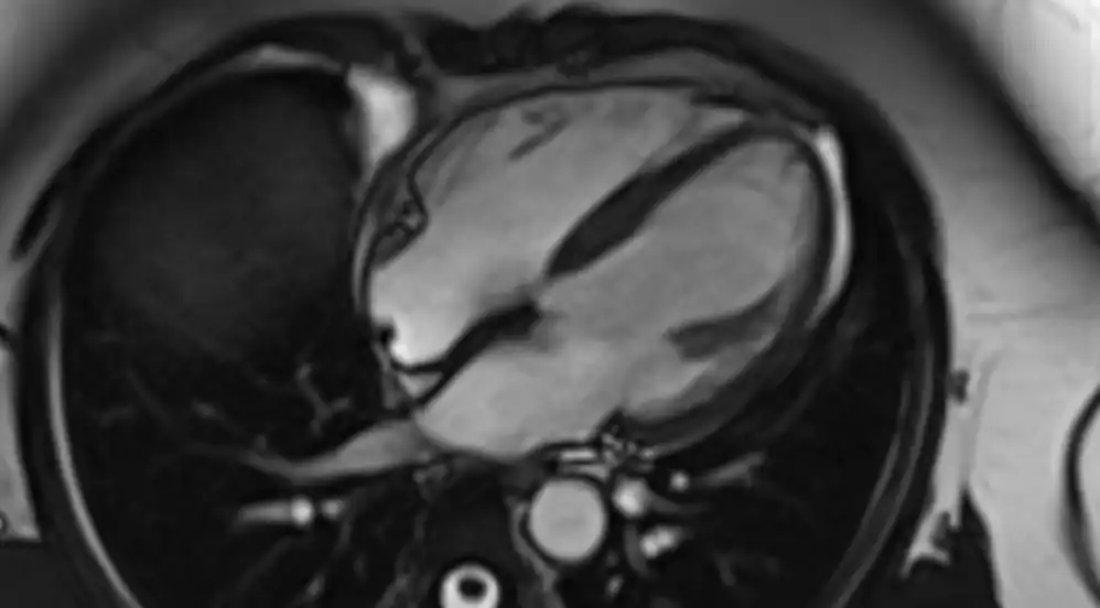
Your heart tells a story and sometimes, seeing it in detail is the only way to truly understand what’s happening inside. A cardiac MRI offers a clear, 3D view of your heart’s structure and function, revealing insights that other tests can’t always show.
A cardiac MRI uses magnets and radio waves to create detailed images of the heart, its valves, chambers, and blood vessels, showing both structure and function. It is a non-invasive test used to diagnose and monitor a wide range of heart conditions such as heart muscle damage, heart failure, congenital heart disease, and tumors—often without using radiation. During the scan, which can last 45 to 90 minutes, patients lie inside a large, tunnel-shaped machine and may be asked to hold their breath at times.
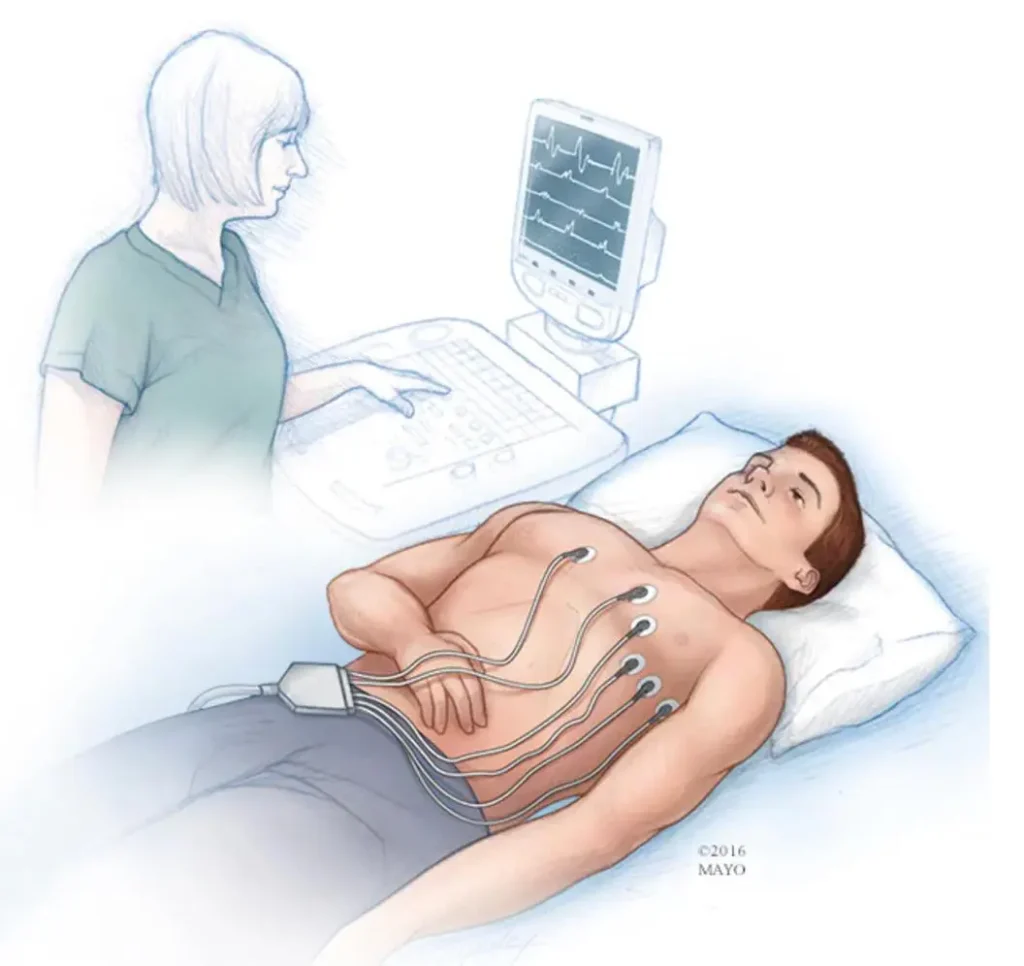
You will lie on a table that slides into a tunnel-shaped MRI scanner. Electrocardiogram (ECG) stickers will be attached to your chest to monitor your heartbeat, and the radiographer may shave a small patch of chest hair for better adhesion.
The machine uses strong magnetic fields and radio waves to take pictures. You will need to lie still to avoid blurred images.
You may be asked to hold your breath for short periods (around 15–20 seconds) to get clearer pictures.
The machine can be very loud, but you will likely be offered earplugs or headphones to listen to music. If you are claustrophobic, talk to your doctor beforehand, as you may be offered a mild sedative.
In some cases, a contrast dye, often containing gadolinium, may be injected through a vein in your arm to provide more detailed images of scar tissue or blood flow.
A cardiac MRI typically takes 45 to 90 minutes, depending on the complexity and the number of images required.
Inform the clinical team before the scan if you have any metallic implants, as the strong magnet can pose a risk.
If you are concerned about confined spaces, speak with your doctor about a sedative before the appointment.
Not every patient with heart concerns needs an MRI, but we may recommend it to:
Often described as a squeezing or heavy feeling, commonly linked to angina or blocked arteries.
Identify areas of the heart muscle affected by previous heart attacks or inflammation.
Check for narrowing or leakage that affects blood flow.
You may be referred for a cardiac MRI if you experience unexplained chest pain, breathlessness, or abnormal results from other heart tests.
When other tests can’t identify the cause.
To check for permanent damage or scarring.
To examine the heart muscle’s thickness and function.
To assess how blood flows through the heart.
To identify myocarditis or pericarditis.
Comprehensive heart care for patients of all ages, addressing chest pain, palpitations, breathlessness, and other cardiac concerns.
Clear answers to common questions about appointments, investigations and what to expect at your first visit.
Dr Badiani manages a wide range of adult cardiology problems including chest pain, breathlessness, palpitations, syncope, heart failure and valve disorders, along with blood pressure and cholesterol management.
You may have an ECG and/or echocardiography arranged. If needed, further tests such as stress echocardiography, ambulatory monitoring or blood tests will be organised promptly.
Appointments require booking; please use the clinic booking portal or contact the clinic to discuss referral or self-referral options.
Consultations are provided in English and Gujarati; please inform reception if you need language support.