
Navigating the world of cardiac diagnostics can be a complex journey, filled with medical terminology that may seem daunting. Two of the most common, yet often confused, procedures are the electrocardiogram (ECG) and the echocardiogram. While both are crucial in assessing heart health, they provide different, yet complementary, information. Understanding the distinction between these two tests is vital for patients seeking to comprehend their cardiac care. This article, with insights from esteemed cardiologist Dr Sveeta Badiani, will demystify these procedures, exploring their unique functions, processes, and the specific cardiac conditions they help to diagnose.
Unveiling the Heart’s Electrical Symphony: The Electrocardiogram (ECG)
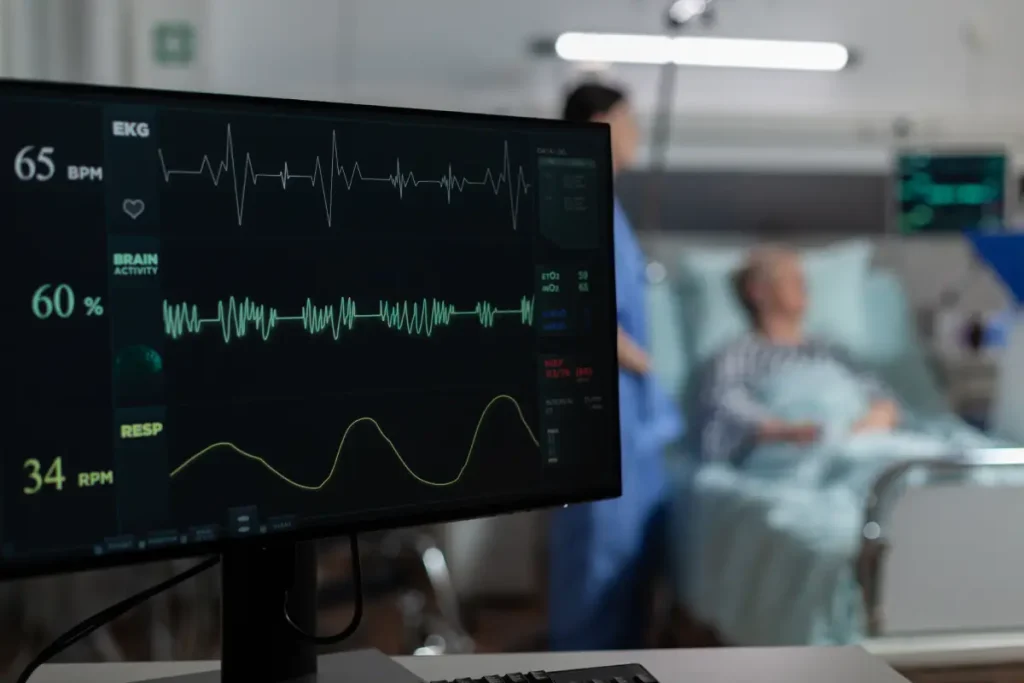
Crucial first line test to investigate heart health. An electrocardiogram, often abbreviated as an ECG or EKG, is a foundational tool in cardiology. It is a non-invasive and painless test that records the electrical activity of the heart. Think of it as listening to the heart’s electrical symphony. Every heartbeat is triggered by an electrical impulse, and the ECG captures these signals, translating them into a visual representation on a monitor or paper. This allows a cardiologist, such as Dr Sveeta Badiani, to analyse the heart’s rhythm and detect any irregularities.
The ECG Procedure: A Quick and Simple Process
The ECG procedure is remarkably straightforward and quick, often completed within a few minutes. During the test, small, sticky sensors called electrodes are placed on the chest, arms, and legs. These electrodes are connected by wires to a machine that records the heart’s electrical signals. The patient is typically asked to lie still and breathe normally during the recording. The simplicity and speed of the ECG make it an invaluable first-line diagnostic tool in both routine check-ups and emergency situations.
What an ECG Reveals
The information gleaned from an ECG is vast. It can reveal the heart’s rate and rhythm, identifying if it is beating too fast (tachycardia), too slow (bradycardia), or irregularly (arrhythmia). Furthermore, an ECG can provide evidence of a previous heart attack or an ongoing one. It can also suggest if the heart is enlarged or if there is a reduced blood supply to the heart muscle. Dr Sveeta Badiani often uses the ECG as a starting point to assess a patient’s cardiac health, guiding further diagnostic steps if necessary.
Types of ECG
While the standard resting ECG is the most common, there are other types that may be used depending on the clinical situation. A stress ECG, for example, is performed while the patient is exercising on a treadmill or stationary bicycle. This helps to assess how the heart responds to physical stress and can be particularly useful in diagnosing coronary artery disease. For patients who experience intermittent symptoms, a Holter monitor may be recommended. This is a small, portable ECG device that is worn for 24 to 48 hours, continuously recording the heart’s electrical activity during normal daily activities. An event monitor is another portable option, which is worn for a longer period and is activated by the patient when they experience symptoms.
A Window into the Heart’s Structure: The Echocardiogram
Examines the conduction system of the heart. While the ECG provides an electrical map of the heart, the echocardiogram offers a detailed, moving picture of the heart’s structure and function. An echocardiogram is a type of ultrasound scan specifically for the heart. It uses high-frequency sound waves to create images of the heart’s chambers, valves, and the major blood vessels connected to it. This allows a cardiologist to observe the heart in real-time as it beats and pumps blood.
The Echocardiogram Procedure: A Detailed Examination
An echocardiogram is also a non-invasive and painless procedure, though it takes longer than an ECG, typically lasting between 30 to 60 minutes. During the test, a small, handheld device called a transducer is coated with a special gel and moved across the chest. The transducer emits sound waves that bounce off the heart, and the returning echoes are converted into images on a screen. This provides a wealth of information about the heart’s size, shape, and how well its chambers and valves are working. Dr Sveeta Badiani may recommend an echocardiogram to investigate symptoms such as shortness of breath or to follow up on findings from an ECG.
What an Echocardiogram Reveals
The echocardiogram provides a comprehensive assessment of the heart’s anatomy and function. It can detect problems with the heart’s valves, such as narrowing (stenosis) or leaking (regurgitation). It can also identify abnormalities in the heart’s muscle, such as thickening or weakness. An echocardiogram is also used to measure the heart’s pumping strength, known as the ejection fraction. This is a crucial indicator of overall heart function. For patients with known heart conditions, Dr Sveeta Badiani may use echocardiograms to monitor the progression of the disease and the effectiveness of treatment.
Types of Echocardiogram
Similar to the ECG, there are different types of echocardiograms. The most common is the transthoracic echocardiogram (TTE), where the transducer is placed on the chest wall. If a more detailed image is required, a transoesophageal echocardiogram (TOE) may be performed. In this procedure, the transducer is passed down the oesophagus, which lies directly behind the heart, providing a clearer view. A stress echocardiogram combines an echocardiogram with a stress test, allowing the cardiologist to see how the heart functions under physical strain. A fetal echocardiogram is a specialised ultrasound performed during pregnancy to evaluate the heart of an unborn baby.
ECG vs. Echocardiogram: A Tale of Two Tests
To summarise the key differences, the ECG and echocardiogram are two distinct yet complementary cardiac investigations. The ECG is an electrical test, while the echocardiogram is a structural and functional test. The ECG is quick and provides information about the heart’s rhythm, while the echocardiogram is more detailed and provides images of the heart’s structure and how it is pumping.
Dr Sveeta Badiani emphasizes that these tests are not mutually exclusive. In fact, they are often used in conjunction to provide a comprehensive picture of a patient’s cardiac health. An abnormal ECG may prompt an echocardiogram to investigate the underlying cause, and an echocardiogram may be used to monitor a condition that was initially detected by an ECG. The choice of test, or combination of tests, will always be tailored to the individual patient’s needs and clinical presentation.
Frequently Asked Questions (FAQs)
Are ECG and echocardiogram tests painful?
No, both the ECG and the standard transthoracic echocardiogram are painless and non-invasive procedures. You may feel a slight coolness from the gel used during the echocardiogram, but there is no discomfort involved. Dr Sveeta Badiani ensures that all procedures are carried out with the utmost patient comfort in mind. For a transoesophageal echocardiogram, a local anaesthetic spray is used to numb the throat, and a sedative may be given to help you relax.
How long do the results of an ECG and echocardiogram take?
The results of an ECG are often available immediately, as the machine records the heart’s electrical activity in real-time. The results of an echocardiogram may take a little longer to be fully interpreted, as the images need to be carefully analysed by a cardiologist. Dr Sveeta Badiani will discuss the results with you at your follow-up appointment, explaining the findings in a clear and understandable manner.
Do I need to prepare for an ECG or echocardiogram?
There is no special preparation required for a standard ECG or transthoracic echocardiogram. For a transoesophageal echocardiogram, you will be asked to avoid eating or drinking for several hours before the procedure. For a stress ECG or stress echocardiogram, you may be advised to wear comfortable clothing and shoes suitable for exercise. Dr Sveeta Badiani’s team will provide you with all the necessary instructions before your appointment.
Can an ECG or echocardiogram detect all heart problems?
While the ECG and echocardiogram are powerful diagnostic tools, they may not detect all heart problems. Some conditions, such as coronary artery disease, may require further investigation with other tests, such as a stress test, cardiac MRI, or coronary angiogram. Dr Sveeta Badiani will recommend the most appropriate diagnostic pathway based on your individual symptoms, risk factors, and the results of your initial tests.
How often should I have an ECG or echocardiogram?
The frequency of these tests depends on your individual health status and risk factors. If you have a known heart condition, Dr Sveeta Badiani may recommend regular monitoring with ECGs or echocardiograms to track the progression of the disease and the effectiveness of treatment. For individuals without any known heart problems, the need for these tests will be determined on a case-by-case basis during a comprehensive cardiac consultation.
Conclusion
In the intricate world of cardiac diagnostics, both the electrocardiogram and the echocardiogram stand as indispensable tools. While the ECG offers a rapid and invaluable assessment of the heart’s electrical system, the echocardiogram provides a detailed and dynamic view of its mechanical function.
By understanding the unique roles of these procedures, patients can engage more effectively in their own healthcare. A collaborative approach, with open communication between the patient and a trusted cardiologist like Dr Sveeta Badiani, is the cornerstone of effective cardiac care. It is through this partnership that the most appropriate diagnostic and treatment strategies can be formulated, leading to the best possible outcomes for heart health.